Plenary Speaker:
Professor Mu-Ming Poo
Short biography
Dr. Mu-ming Poo was born in China, received his B.S. in physics from Tsinghua University in Taiwan (1970), and Ph.D in biophysics from Johns Hopkins University, Baltimore (1974). Following postdoctoral training at Purdue University, he was appointed as Assistant Professor in Physiology at the University of California, Irvine in 1976, promoted to Associate Professor in 1979 and Professor in 1983. In 1985, he moved to Yale University School of Medicine, where he served as Research Professor in Molecular Neurobiology. During 1988-95, he was Professor of Biological Sciences at Columbia University. During 1995-2000, he held the Stephen W. Kuffler Chair in Neurobiology at University of California at San Diego and in 2000, he moved to the University of California, Berkeley. He was the Paul Licht Distinguished Professor in the Department of Molecular and Cell Biology in 2006-2013 and the head of the neurobiology division in 2001-2006. Since 1999, he has served as the founding Director of the Institute of Neuroscience, Chinese Academy of Sciences and the Head of the Laboratory of Neural Plasticity. He is the member of Chinese Academy of Science (foreign member), US National Academy of Sciences and Academia Sinica. In 2014, he was appointed as the director of the CAS Center for Excellence in Brain Science. Dr. Poo’s Research Interests focus on cellular and molecular mechanisms underlying axon guidance, synaptogenesis and synaptic plasticity. He has received the following honors: AAAS Fellow (2001), Ameritec Prize (2001), Ray Wu Society Award (2002), Docteur Honoris Causa of Ecole Normale Supérieure, Paris (2003), National Prize for International Cooperation of China (2005), Qiushi Excellent Scientist Award (2010), Outstanding Science and Technology Achievement Prize of the Chinese Academy of Sciences (Research Group, 2011) , Honorary Doctoral Degree from the Hong Kong University of Science and Technology (2014), Gruber Neuroscience Prize (2016).
Presentation title and abstract
Pattern Recognition by the Brain: Neural Circuit Mechanisms
The brain acquires the ability of pattern recognition through learning. Understanding neural circuit mechanisms underlying learning and memory is essential for understanding how the brain recognizes patterns. Much progress has been made in this area of neuroscience during the past decades. In this lecture, I will summarize three distinct features of neural circuits that provide the basis of learning and memory of neural information. First, the architecture of the neural circuits is continuously modified by experience. This process of experience-induced sculpting (pruning) of connections is most prominent early in development and decreases gradually to a much more limited extent in the adult brain. Second, the efficacy of synaptic transmission could be modified by neural activities associated with experience, in a manner that depends on the pattern (frequency and timing) of spikes in the pre- and postsynaptic neurons. This activity-induced circuit alteration in the form of long-term potentiation (LTP) and long-term depression (LTD) of existing synaptic connections is the predominate mechanism underlying learning and memory of the adult brain. Third, learning and memory of information containing multiple modalities, e.g., visual, auditory, and tactile signals, involves processing of each type of signals by different circuits for different modalities, as well as binding of processed multimodal signals through mechanisms that remain to be elucidated. Two potential mechanisms for binding of multimodal signals will be discussed: binding of signals through converging connections to circuits specialized for integration of multimodal signals, and binding of signals through correlated firing of neuronal assemblies that are established in circuits for processing signals of different modalities. Incorporation of these features into artificial neural networks may help to achieve more efficient pattern recognition, especially for recognition of time-varying multimodal signals.
Keynote-Special Speaker:
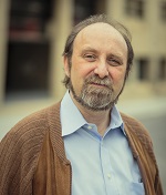
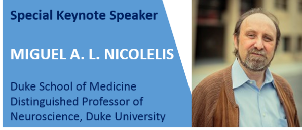
Professor MIGUEL A. L. NICOLELIS
Short biography
In creating the field of neuroengineering, Miguel A.L. Nicolelis’ pioneering work on brain-machine interfaces has completely changed people’s perception of what brains can do and how such research can be rapidly applied to help humans. Nicolelis demonstrated that humans can use raw brain activity to directly communicate with mechanical, electronic, and virtual devices in real time and in a closed control loop. He played a key role in the development of a robotic exoskeleton that can help paralyzed individuals to walk. He focused on methods to read a paraplegic person’s brain waves and decode and use them to move hydraulic drivers on the suit. His work has great implications for patients with epilepsy, Parkinson’s disease, and spinal cord injury. An IEEE Member, Nicolelis is the Duke School of Medicine Distinguished Professor of Neuroscience at Duke University, Durham, NC, USA. TAGLINE: Leader in developing neuroprosthetic devices that harness brain activity is revolutionizing therapies for patients suffering from severe levels of paralysis.
Presentation title and abstract: TBD
Keynote Speakers:
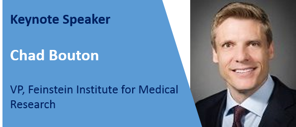
Professor Chad Bouton
Short biography
Prof. Chad Bouton is the VP of Advanced Engineering and the Director of the Center for Bioelectronic Medicine at The Feinstein Institute for Medical Research, the research arm of the Northwell Health System in New York. Professor Bouton formerly served as research leader at Battelle Memorial Institute—the world’s largest independent research and development organization—where he spent 18 years researching and developing biomedical technology. At the Feinstein Institute, he is performing groundbreaking research in neurotechnology to treat paralysis and is developing new technologies to accelerate the field of bioelectronic medicine. Professor Bouton’s pioneering work, allowing a paralyzed person for the first time to regain movement using a brain implant, has been featured on 60 Minutes, CBS, and presented at TEDx. He holds over 70 patents worldwide and his technologies have been awarded three R&D 100 Awards and he was recognized by the US Congress for his work in the medical device field. He has been named Inventor of the Year and Distinguished Inventor by Battelle, and was selected by the National Academy of Engineering in 2011 to attend the Frontiers in Engineering Symposium.
Presentation title and abstract
Restoring Movement after Paralysis and the Future of Bioelectronic Medicine
Millions of people worldwide currently suffer from diseases and injuries leading to severe paralysis. Bioelectronic technology is now being developed to restore movement by decoding and re-routing neural signals around damaged or degenerated neural pathways. It has previously been shown that signals recorded in the brain can be decoded and linked to assistive technology and robotic devices. In non-human primates, these signals have also been used to activate chemically paralyzed arm muscles. More recently, it has been shown signals recorded within the brain can be linked in real-time to muscle activation to restore movement in a paralyzed human. Functional movement and rhythmic movement have now both been demonstrated, with the potential to have a positive impact on the quality of life for paralyzed patients. Bioelectronic medicine research has opened many new doors to ways for treating not only spinal cord injury, but potentially stroke and brain injury as well, leading to many potential treatment options for patients worldwide in the future.
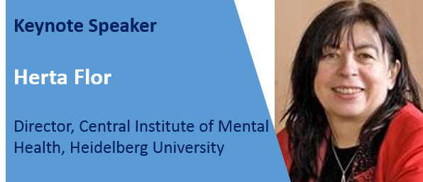
Professor Herta Flor
Short biography
Herta Flor is Full Professor of Neuropsychology and Clinical Psychology and Scientific Director of the Department of Cognitive and Clinical Neuroscience at the Central Institute of Mental Health, Medical Faculty Mannheim, Heidelberg University, Germany. Her research focuses on the role of neuronal plasticity, learning and memory in chronic pain, anxiety and personality disorders, substance abuse and on novel training-based therapies. She was spokesperson of Collaborative Research Center 636: “Learning, memory and brain plasticity: Implications for psychopathology” funded by the German Research Foundation. She is a member of the German Academy of Sciences Leopoldina and the Academia Europaea and holds an honorary doctoral degree from the Free University Amsterdam. She received numerous awards, among them the lifetime achievement award of the German Psychological Association and the Max-Planck Research Award. Herta Flor is the recipient of more than 60 research grants, among them a European Research Council Advanced Grant and a Koselleck award of the German Research Foundation. She is the author of more than 450 scholarly papers.
Presentation title and abstract
Brain circuits involved in phantom limb pain: Implications for treatment
Phantom limb pain is a frequent sequel of an amputation occurring in up to 80% of the amputee population. Peripheral factors such as local changes at the residual limb, alterations in severed nerves and associated dorsal root ganglia, as well as central changes such as spinal and supraspinal plastic changes including structural and functional changes have been examined. Moreover, input from the amputated limb has been discussed as a crucial factor. New insights about central changes have also come from studies on somatosensory illusions and altered body perception. These hypotheses have led to new behavioral and pharmacological treatment option that target maladaptive plasticity and learning and sensory incongruence phenomena. The treatments include prosthesis use, discrimination training, mirror treatment, video feedback, imagery, virtual reality applications, brain-computer interfaces as well as combinations of these treatments with pharmacological interventions. We will provide a critical overview of these recent developments on sources and treatments of phantom limb pain and discuss the need for causal experimental designs.
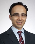
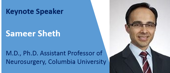
Professor Sameer Sheth
Short biography
Sameer Sheth, M.D., Ph.D. Assistant Professor of Neurosurgery, Columbia University.
Presentation title and abstract
Neurophysiology of Cognitive Control in Human Prefrontal Cortex
In our day-to-day lives, we are constantly faced with decisions. For example, when approaching an intersection in which the traffic light turns yellow, we must make an optimal decision about whether to hit the brake or accelerator. Our brain’s decision-making circuitry must incorporate relevant information (speed, distance to the intersection, presence of police, etc.), ignore irrelevant information (children arguing in the back seat), factor in long-term goals (avoid a traffic violation), and modify future behavior based on previous outcomes. The neurocognitive processes that permit such controlled decision-making can be subsumed under the term “cognitive control,” which refers to the allocation of mental resources to optimize the performance of goal-driven behavior. The prefrontal cortex (PFC) is known to play an important role in cognitive control. We have studied these processes in the human brain using the opportunities afforded by neurosurgical procedures requiring intracranial neurophysiological recordings, such as deep brain stimulation or epilepsy surgery. In these procedures, we typically have to place electrodes in a variety of brain regions for clinical purposes. Consenting patients can then engage in behavioral tasks that allow us to study components of the decision-making process. Using this paradigm, we have recorded from two critical regions in the cognitive control network, the dorsal anterior cingulate cortex (dACC) and dorsolateral PFC (dlPFC). We find that individual dACC neurons encode specific aspects of stimulus parameters that are relevant for the decision. These neurons’ firing rate also encodes an estimate of the amount of cognitive effort required to perform the task. Analysis of population activity (local field potentials, LFP) reveals that this information is transmitted from dACC to dlPFC using a “carrier signal” in the form of oscillatory activity in the theta (4-7 Hz) range. dACC neurons conveying relevant information synchronize their firing to theta oscillations, which carry the information over the relatively large expanse to dlPFC. A large fraction of dlPFC neurons, in turn, synchronize their firing to the theta signal. This spike-LFP coherence is a likely mechanism for coordinating information transfer between individual neurons and larger neural populations across brain regions. Precise temporal coordination of these signals between these structures allows the decision-making circuitry to optimize the selection and timing of our responses, enabling us to seamlessly execute well-controlled behaviors.
The latest update on March 23th, 2017

